Home

bibliotecă adiacent Catâr electron microscope ribosome Inactiv snorkel moral

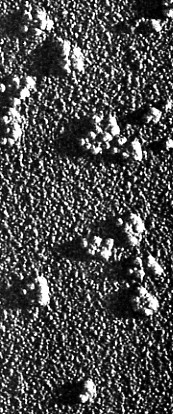
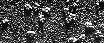
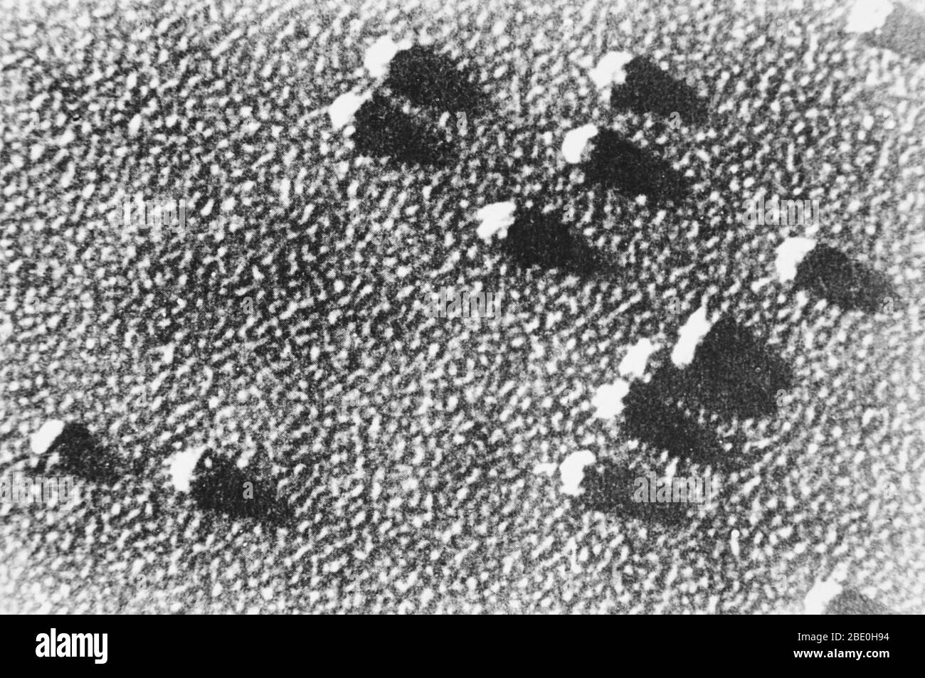
Scanning electron micrograph (SEM) showing ribosome particles. The ribosome  is a complex molecular machine, found within all living cells, that serves  as the site of biological protein synthesis (translation). Ribosomes link  amino
Scanning electron micrograph (SEM) showing ribosome particles. The ribosome is a complex molecular machine, found within all living cells, that serves as the site of biological protein synthesis (translation). Ribosomes link amino

Ribosome | British Society for Cell Biology

ELECTRON MICROSCOPE STUDY OF MITOCHONDRIAL 60S AND CYTOPLASMIC 80S RIBOSOMES  FROM LOCUSTA MIGRATORIA
ELECTRON MICROSCOPE STUDY OF MITOCHONDRIAL 60S AND CYTOPLASMIC 80S RIBOSOMES FROM LOCUSTA MIGRATORIA

The Ribosome
The Ribosome

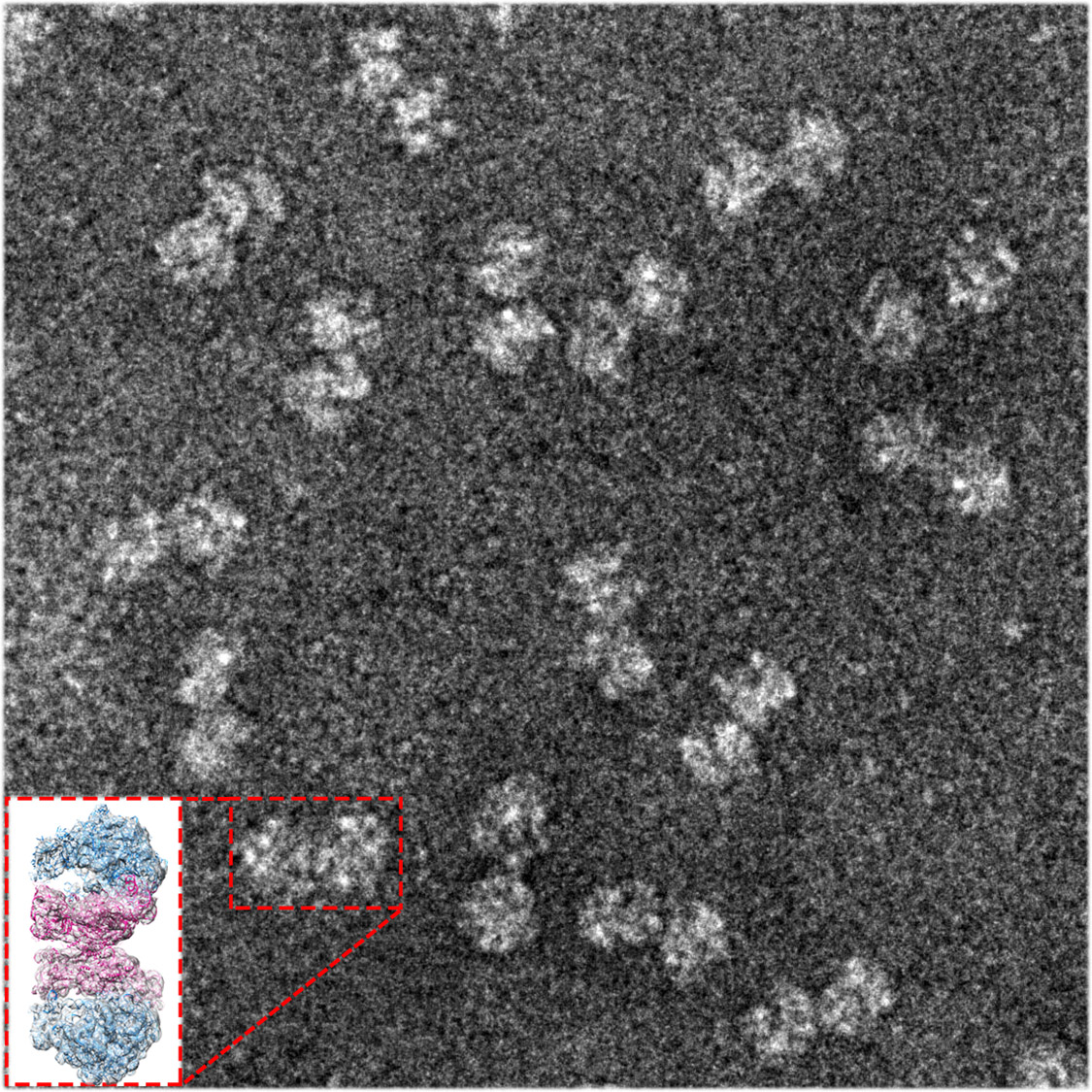
Cryo electron micrograph depicting the distribution of 80S particles at...  | Download Scientific Diagram
Cryo electron micrograph depicting the distribution of 80S particles at... | Download Scientific Diagram

Ribosomes | Celebrate Cytochemistry | Gwen V. Childs, Ph.D.

Electron microscopy led to the first identification of what would later be  known as ribosomes. | Structural biology, Microscopy, Electrons
Electron microscopy led to the first identification of what would later be known as ribosomes. | Structural biology, Microscopy, Electrons

Electron Micrographs

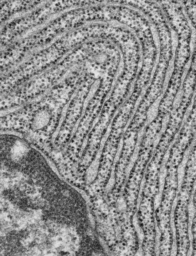
RER - Dr.Jastrow's EM-Atlas

The Protein that Puts Staphylococcus Ribosomes to Sleep - Weizmann

2.3.3 Identify structures from electron micrographs of liver cells - YouTube

Electron-Microscopy-Milestone

ELECTRON MICROSCOPE STUDY OF MITOCHONDRIAL 60S AND CYTOPLASMIC 80S RIBOSOMES  FROM LOCUSTA MIGRATORIA
ELECTRON MICROSCOPE STUDY OF MITOCHONDRIAL 60S AND CYTOPLASMIC 80S RIBOSOMES FROM LOCUSTA MIGRATORIA

Ribosomes, Mitochondria, and Peroxisomes | Biology for Majors I

Ribosomes
Ribosomes

Ribosomes, Transcription, Translation | Learn Science at Scitable

LON-CAPA Botany online: Intracellular Structures - Electron Microscopic  Images
LON-CAPA Botany online: Intracellular Structures - Electron Microscopic Images

Ribosome Structure Determined by Electron Microscopy of Escherichia coli  Small Subunits, Large Subunits and Monomeric Ribosomes
Ribosome Structure Determined by Electron Microscopy of Escherichia coli Small Subunits, Large Subunits and Monomeric Ribosomes
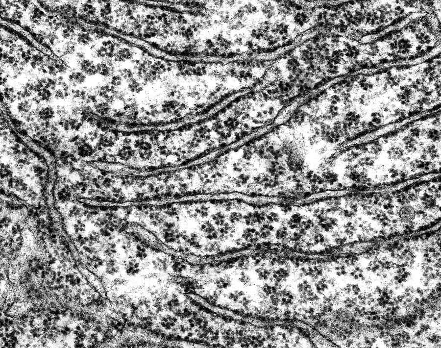
3.4: Ribosomes - Biology LibreTexts

Ribosomes | Highbrow

Rough Endoplasmic Reticulum With Ribosomes Photograph by Dennis Kunkel  Microscopy/science Photo Library - Pixels
Rough Endoplasmic Reticulum With Ribosomes Photograph by Dennis Kunkel Microscopy/science Photo Library - Pixels

Nucleolus - Wikipedia

ribosome Dr.Jastrow's EM-Atlas

File:A-general-mechanism-of-ribosome -dimerization-revealed-by-single-particle-cryo-electron-microscopy-41467  2017 718 MOESM2 ESM.ogv - Wikimedia Commons
File:A-general-mechanism-of-ribosome -dimerization-revealed-by-single-particle-cryo-electron-microscopy-41467 2017 718 MOESM2 ESM.ogv - Wikimedia Commons

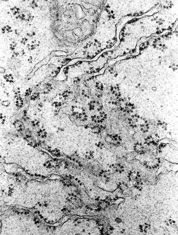
Electron microscopy images of ribosomal dimers present in the 110S peak...  | Download Scientific Diagram
Electron microscopy images of ribosomal dimers present in the 110S peak... | Download Scientific Diagram

Electron micrographs of reticulocyte ribosomes. The ribosomes were... |  Download Scientific Diagram
Electron micrographs of reticulocyte ribosomes. The ribosomes were... | Download Scientific Diagram

Can mRNA molecules be seen with an electron microscope? - Quora

Photograph | Cell Structure, Ribosomes, TEM | Science Source Images