Home

extaz in afara serviciului victorie lamina propria si musculara în mijlocul pustiului Unde Alice

Solved LAYERS OF THE DIGESTIVE TRACT WALL net V Mucosa | Chegg.com

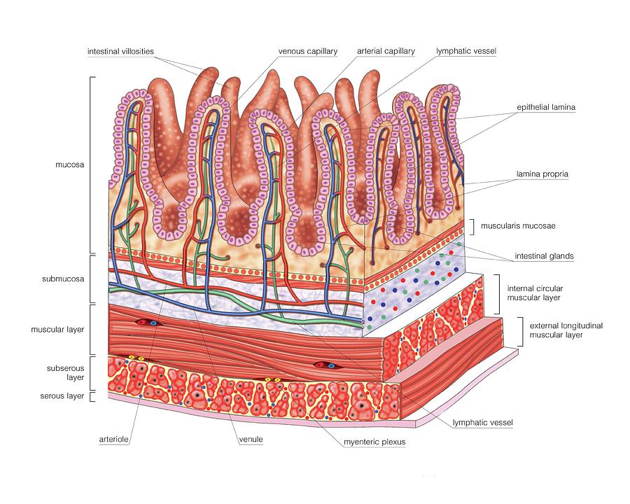
Structure Of Intestinal Tract Poster by Asklepios Medical Atlas - Fine Art  America
Structure Of Intestinal Tract Poster by Asklepios Medical Atlas - Fine Art America

Solved In order to remove a tumor in the muscular tunic of | Chegg.com

Neoplasmul colo-rectal – Spitalul Universitar de Urgenţă Militar Central  Dr. Carol Davila
Neoplasmul colo-rectal – Spitalul Universitar de Urgenţă Militar Central Dr. Carol Davila

Small Intestine by Asklepios Medical Atlas

Small intestine cross section showing mucosa, submucosa, lamina propria,  muscular layer, Stock Photo, Picture And Rights Managed Image. Pic.  VD7-2972289 | agefotostock
Small intestine cross section showing mucosa, submucosa, lamina propria, muscular layer, Stock Photo, Picture And Rights Managed Image. Pic. VD7-2972289 | agefotostock

Telocytes constitute a widespread interstitial meshwork in the lamina  propria and underlying striated muscle of human tongue | Scientific Reports
Telocytes constitute a widespread interstitial meshwork in the lamina propria and underlying striated muscle of human tongue | Scientific Reports

Lamina propria - Wikipedia

05.Tubul digestiv_2018_site.pptx

Small intestine cross section showing mucosa, submucosa, lamina propria,  muscular layer, Stock Photo, Picture And Rights Managed Image. Pic.  VD7-2972289 | agefotostock
Small intestine cross section showing mucosa, submucosa, lamina propria, muscular layer, Stock Photo, Picture And Rights Managed Image. Pic. VD7-2972289 | agefotostock

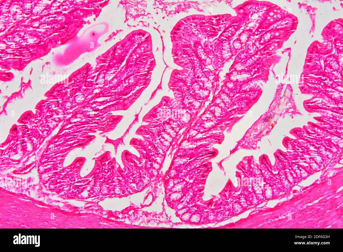
Rectum (large intestine) showing muscular layer, lamina propria, submucosa,  mucosa, epithelium, villi and intestinal glands. Optical microscope X100  Stock Photo - Alamy
Rectum (large intestine) showing muscular layer, lamina propria, submucosa, mucosa, epithelium, villi and intestinal glands. Optical microscope X100 Stock Photo - Alamy

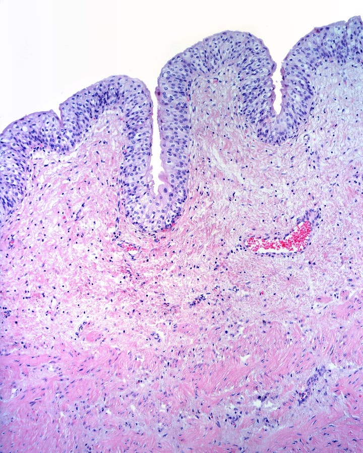
Urinary bladder mucosa stock photo. Image of histology - 117239608

Understanding Barrett's Esophagus

Neoplasmul colo-rectal – Spitalul Universitar de Urgenţă Militar Central  Dr. Carol Davila
Neoplasmul colo-rectal – Spitalul Universitar de Urgenţă Militar Central Dr. Carol Davila

Superficial, intermediate, and deep layers of lamina propria. A)... |  Download Scientific Diagram
Superficial, intermediate, and deep layers of lamina propria. A)... | Download Scientific Diagram

Enteric Muscular System Gut Wall Small Intestine Outline Diagram Labeled  Stock Vector Image by ©VectorMine #596447086
Enteric Muscular System Gut Wall Small Intestine Outline Diagram Labeled Stock Vector Image by ©VectorMine #596447086
Colonic Lamina Propria - an overview | ScienceDirect Topics

Rectum (large intestine) showing muscular layer, lamina propria, submucosa,  mucosa, epithelium, villi and intestinal glands. Optical microscope X100  Stock Photo - Alamy
Rectum (large intestine) showing muscular layer, lamina propria, submucosa, mucosa, epithelium, villi and intestinal glands. Optical microscope X100 Stock Photo - Alamy

Lamina propria: The functional center of the bladder? - Andersson - 2014 -  Neurourology and Urodynamics - Wiley Online Library
Lamina propria: The functional center of the bladder? - Andersson - 2014 - Neurourology and Urodynamics - Wiley Online Library

Small and Large intestine Histology Flashcards | Quizlet

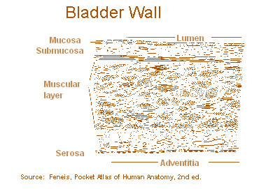
SEER Training: Layers of the Bladder Wall

The urethral wall was measured in the lamina propria to the outer limit...  | Download Scientific Diagram
The urethral wall was measured in the lamina propria to the outer limit... | Download Scientific Diagram

Layers of the esophagus stock image. Image of microscope - 117240065

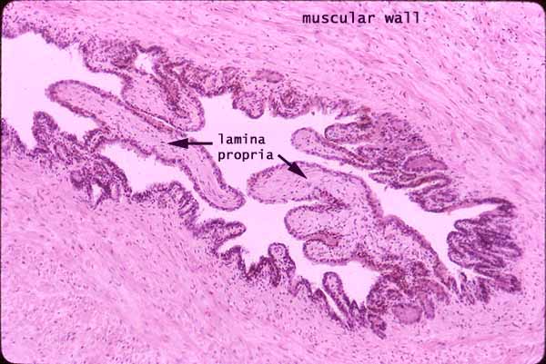
Histology at SIU
Histology at SIU

Rectum (large intestine) showing adventitia, muscular layer, lamina propria,  submucosa, mucosa, epithelium, villi and intestinal glands. Optical Stock  Photo - Alamy
Rectum (large intestine) showing adventitia, muscular layer, lamina propria, submucosa, mucosa, epithelium, villi and intestinal glands. Optical Stock Photo - Alamy