Home

Al treilea tejghea Al patrulea oct microscope pașaport Slăbi sol

Microscope integrated optical coherence tomography system combined with  augmented reality
Microscope integrated optical coherence tomography system combined with augmented reality

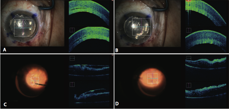
Real-time OCT images of a microscope-integrated spectral domain OCT... |  Download Scientific Diagram
Real-time OCT images of a microscope-integrated spectral domain OCT... | Download Scientific Diagram
Using microscope-integrated intraoperative OCT in VR surgery

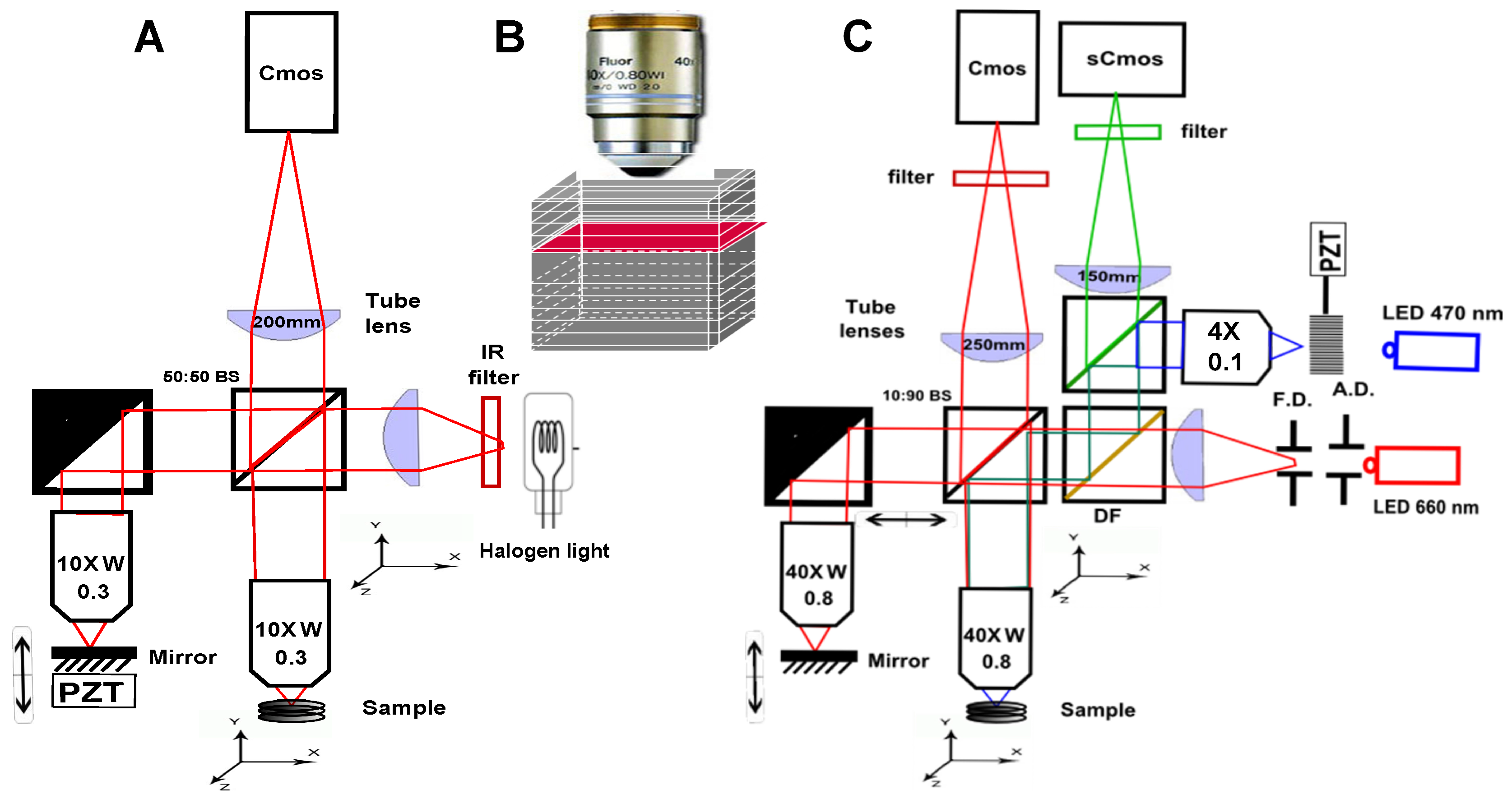
Applied Sciences | Free Full-Text | Full-Field Optical Coherence Tomography  as a Diagnosis Tool: Recent Progress with Multimodal Imaging
Applied Sciences | Free Full-Text | Full-Field Optical Coherence Tomography as a Diagnosis Tool: Recent Progress with Multimodal Imaging

Fig. 8.2, [Optical coherence tomography (OCT) for...]. - High Resolution  Imaging in Microscopy and Ophthalmology - NCBI Bookshelf
Fig. 8.2, [Optical coherence tomography (OCT) for...]. - High Resolution Imaging in Microscopy and Ophthalmology - NCBI Bookshelf

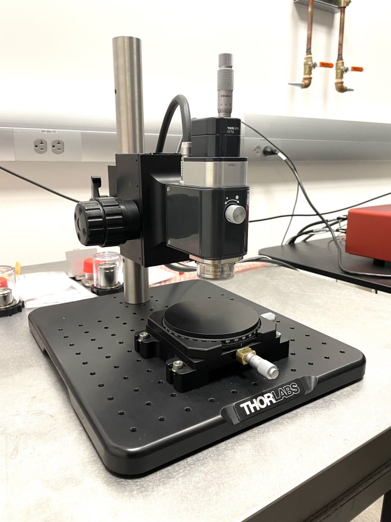
Optical Coherence Tomography (Thorlabs) - McMaster Centre for Advanced  Light Microscopy (CALM)
Optical Coherence Tomography (Thorlabs) - McMaster Centre for Advanced Light Microscopy (CALM)

Microscope-Integrated Optical Coherence Tomography - Retina Today

Applied Sciences | Free Full-Text | Optical Coherence Tomography (OCT) for  Time-Resolved Imaging of Alveolar Dynamics in Mechanically Ventilated Rats  | HTML
Applied Sciences | Free Full-Text | Optical Coherence Tomography (OCT) for Time-Resolved Imaging of Alveolar Dynamics in Mechanically Ventilated Rats | HTML

Figure 4 from The use of optical coherence tomography in intraoperative  ophthalmic imaging. | Semantic Scholar
Figure 4 from The use of optical coherence tomography in intraoperative ophthalmic imaging. | Semantic Scholar
Optical coherence tomography - Wikipedia

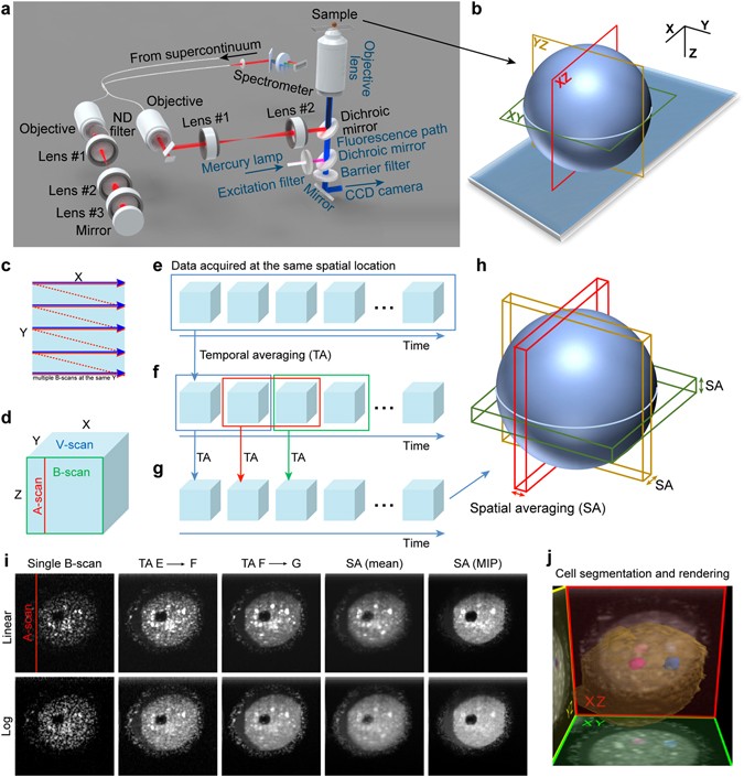
Optical coherence microscopy as a novel, non-invasive method for the 4D  live imaging of early mammalian embryos | Scientific Reports
Optical coherence microscopy as a novel, non-invasive method for the 4D live imaging of early mammalian embryos | Scientific Reports

Introduction to OCT - OBEL

First OCT-guided intraoperative microscope | Institut Català de Retina

Schematic of LC-OCT, combining the principles of OCT and line-scanning... |  Download Scientific Diagram
Schematic of LC-OCT, combining the principles of OCT and line-scanning... | Download Scientific Diagram

Introduction to OCT - OBEL

Lightweight Learning-Based Automatic Segmentation of Subretinal Blebs on  Microscope-Integrated Optical Coherence Tomography Images - American  Journal of Ophthalmology
Lightweight Learning-Based Automatic Segmentation of Subretinal Blebs on Microscope-Integrated Optical Coherence Tomography Images - American Journal of Ophthalmology

Millimeter-scale chip–based supercontinuum generation for optical coherence  tomography | Science Advances
Millimeter-scale chip–based supercontinuum generation for optical coherence tomography | Science Advances

Intrasurgical OCT EnFocus - SurgicalOne

Schematic of the FF-OCT system. BS: beam splitter, OL: microscope... |  Download Scientific Diagram
Schematic of the FF-OCT system. BS: beam splitter, OL: microscope... | Download Scientific Diagram

Time-domain optical coherence tomography with digital holographic microscopy

a) Schematic of the surgical microscope/OCT system; (b) Surgical head... |  Download Scientific Diagram
a) Schematic of the surgical microscope/OCT system; (b) Surgical head... | Download Scientific Diagram

Evolving the surgical microscope | EurekAlert!

Utility of microscope-integrated optical coherence tomography (MIOCT) in  the treatment of myopic macular hole retinal detachment | BMJ Case Reports
Utility of microscope-integrated optical coherence tomography (MIOCT) in the treatment of myopic macular hole retinal detachment | BMJ Case Reports

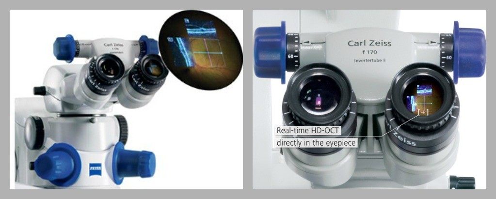
Figure 3 from Microscope-integrated optical coherence tomography:  Single-center experience with the Zeiss Rescan 700 microscope-mounted OCT  system | Semantic Scholar
Figure 3 from Microscope-integrated optical coherence tomography: Single-center experience with the Zeiss Rescan 700 microscope-mounted OCT system | Semantic Scholar